
Ultrasound Imaging
Ultrasound Imaging Customer Case This customer's application involves capturing ultrasound echoes from a medical imaging system, to be used in bedside monitors, defibrillators, intervascular ultrasound, ECG, etc. The live imaging rate is 30 frames per second (fps). Each image frame depicts a full 360-degree circular section. A frame is 240 echoes, so the pulse repeat frequency (PRF) is 7200 Hz, or one echo every 140 us. The customer anticipates signals at 50 MHz with a 50% bandwidth (the passband must extend to at least 75 MHz). The echo duration is 15 us; the signal size is roughly +/- 70 [...]
Ultrasonic Imaging
Ultrasonic Imaging Customer Case The customer is digitizing signals returned from an ultrasonic transducer used in the study of cardiac phenomena. At the moment, a 5 MHz transducer is being used, but the customer wants to upgrade to a 10 MHz transducer in the near future. This transducer will give him better spacial resolution. The customer needs one channel of very fast (100 MSPS), preferably 12-bit A/D conversion which is controlled by an external trigger signal. Bandwidth of the analog front-end of the A/D card or system must be very flat up to 15 or 20 MHz. The application [...]

How Capacitive Measurement Can Help Reduce Packaging Waste
Product packaging is often made of lightweight polymeric foams or plastic sheets and films. These materials are relatively inexpensive, but manufacturers still want to minimize waste and maximize yields. Across high volumes of low-cost materials, even a small per-unit savings can become significant, especially as the price of petroleum – a key ingredient in many polymers – continues to rise. For companies with environmental sustainability efforts, there are other important considerations as well. The excessive use of packaging materials can contribute to environmental pollution, especially with polymers that are slow to degrade. Yet, materials such as plastic sheets and [...]
Ultrasound Tissue Imaging
Ultrasound Tissue Imaging Customer Case This customer is building a system to be used as a test station for examining human tissue for disease. Diseased organs differ from healthy organs in ways that can be measured via the echoes of ultrasound transmissions which are passed through the tissues. The customer's system will build a 3D image of the tissue under examination. In order to obtain a clear ultrasound image of the object being studied, the successive data captures have to be aligned to eliminate a distorted image. Jitter which would result from an asynchronous triggering is not acceptable. The [...]
Ultrasonic Medical Imaging System
Ultrasonic Medical Imaging System Customer Case A customer wants to construct an ultrasonic medical imaging system based on a transducer array. A central ultrasonic generation transducer generates a burst of ultrasonic energy which travels towards a tissue specimen. Echoes reflected off the tissue specimen are simultaneously detected by an array of 48 ultrasonic receiver transducers arranged in a dish-like pattern. The customer must simultaneously digitize all 48 signals and download them to PC RAM as quickly as possible. The customer requires 8 bit vertical resolution and would like to sample at 40 MS/s, but 20 MS/s would be acceptable. [...]
Intra-Vascular Ultrasound
Intra-Vascular Ultrasound Customer Case The customer does intra-vascular diagnostic ultrasound. In this novel technique, an ultrasonic catheter is guided through a patient's blood vessel. Throughout its helical or corkscrew-like path, the ultrasonic transducer-tipped catheter emits bursts of ultrasonic energy. Reflections from the vessel wall return into the transducer and can be used to characterize such features as plaque deposits. The customer wishes to resolve features as small as 3 mm. The amount of time, D t, required for ultrasound to reflect off an interface that is a distance x away and return to the transducer is: D t = [...]
Deep Capture of Medical Sonography Signals
Deep Capture of Medical Sonography Signals Customer Case Medical sonography refers to the use of reflected ultrasonic echoes to image a patient's internal organs. A customer has a commercial medical diagnostic ultrasound system. During its operation, an external probe is placed upon the patient. An ultrasonic transducer within the probe head mechanically scans in a linear frame pattern at a rate of approximately 22 frames/sec. The transducer generates a sharp ultrasonic pulse into the patient and the same transducer detects reflected echoes from various tissue interfaces. In normal operation, the system processes the ultrasonic echoes in a prescribed fashion [...]


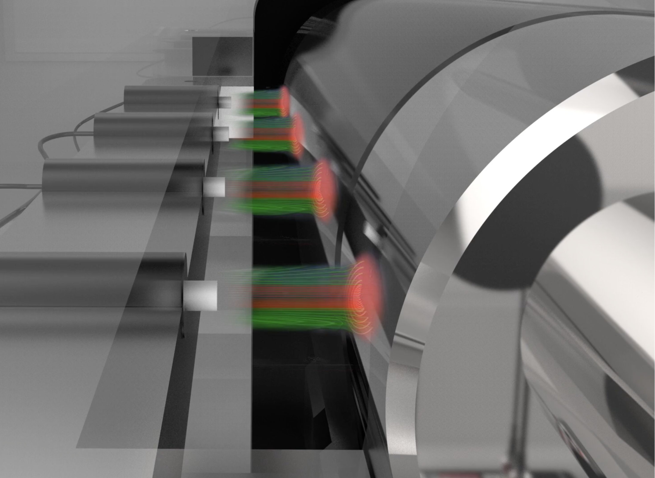
Non-Contact Displacement Sensors for Detection and Measurement
Non-contact displacement sensors are designed to detect or measure physical properties such as linear displacement, thickness, proximity, and distance without physically touching the target. By emitting various forms of energy, such as capacitance, fiber optic, or laser triangulation, these sensors capture highly accurate readings without physically touching the object. This contrasts with traditional contact sensors, which rely on direct mechanical contact to gauge displacement or other physical properties. Benefits of Non-Contact vs. Contact Displacement Sensors Compared to contact sensors, non-contact displacement sensors offer faster response times, higher sampling rates, and minimal interference with the object being measured. They are [...]
Laser Scanning Microscope Interface
Laser Scanning Microscope Interface Customer Case This customer needs to interface a scanning laser microscope to their PC so that image data can be acquired and stored. Data is received from the laser microscope via an optical sensor at 1300 line scans per image. Each scan has an 8 ms duration, but 2/3 of that time is scan time and 1/3 is flyback time (dead time). The customer has determined that a 1 us sample width (or faster) would be adequate. There is also a price vs. performance issue - the customer has a tight budget and wants the [...]
Electron Paramagnetic Resonance Imaging (EPRI) Measuring of Tumor Oxygen Levels
Electron Paramagnetic Resonance Imaging (EPRI) Measuring of Tumor Oxygen Levels Customer Case The customer is improving their Electron Paramagnetic Resonance Imaging (EPRI) system that is used for the diagnosis of tumors. Using this non-invasive method, the concentration of oxygen in living tissues can be determined without the need for biopsies. Tumors that have little or no oxygen content (i.e. hypoxia) also have increased resistance to radio-frequency radiation, making it less effective as a choice for cancer treatment. Identifying those patients with hypoxic tumors allow alternative treatments to be used instead. The requirement is to capture the returned RF signals [...]