
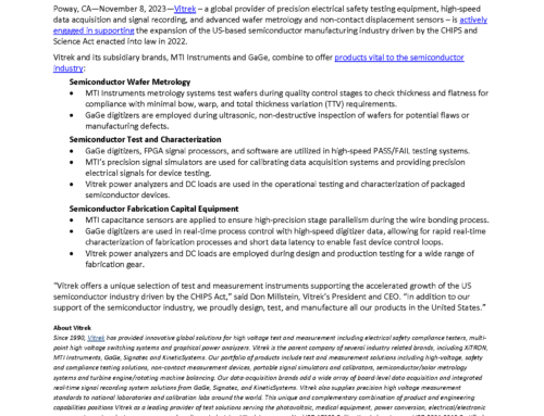
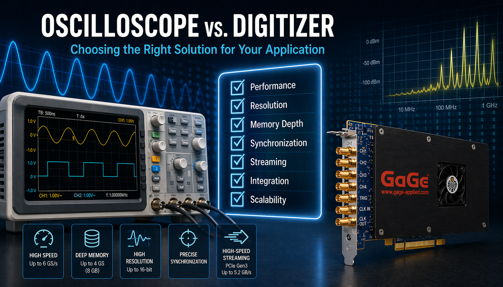
Introduction
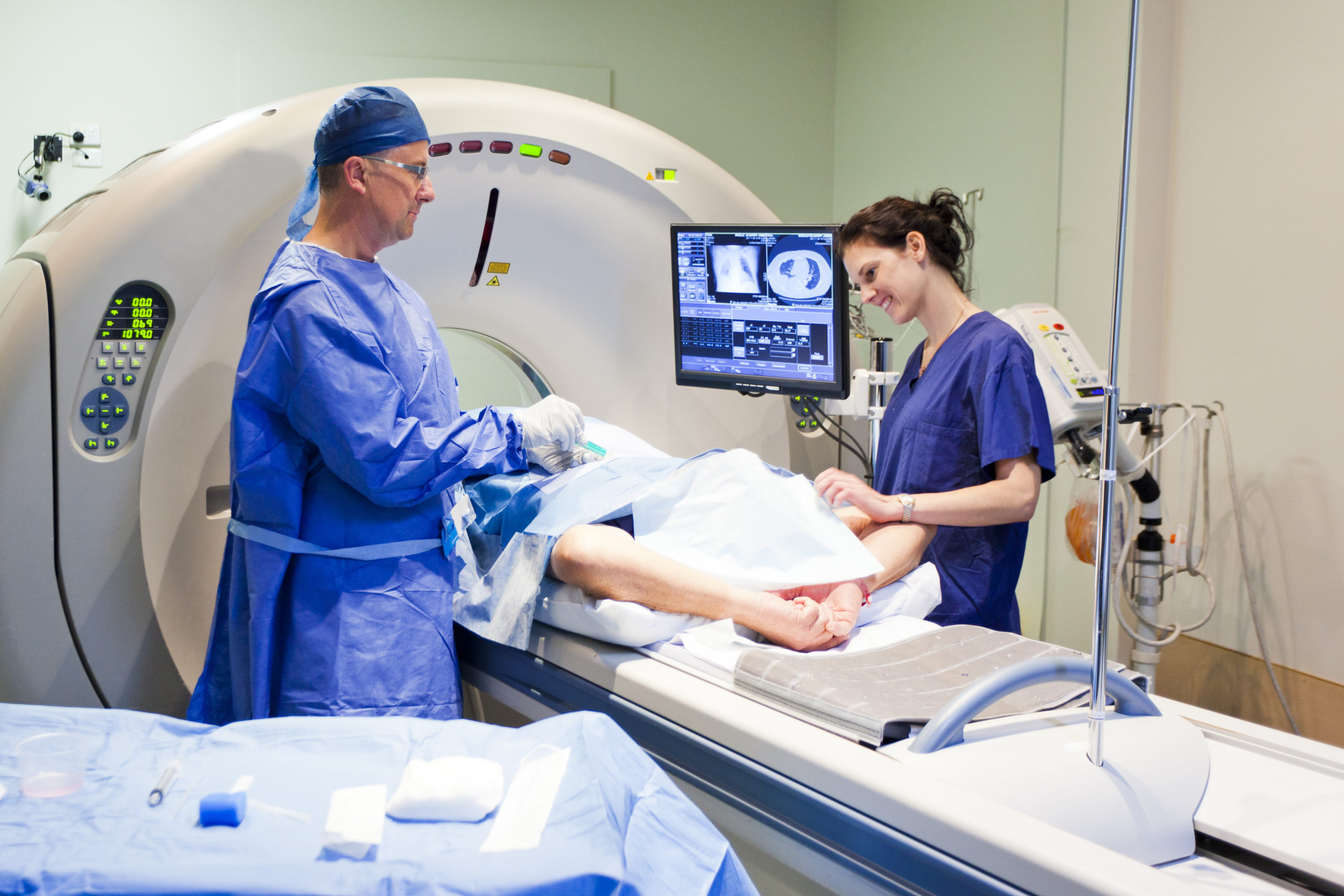
High-speed digitizers are indispensable tools in the medical and pharmaceutical industries, transforming how data is captured, processed, and analyzed. These advanced devices enable precise measurement and real-time monitoring in applications ranging from medical imaging and diagnostics to biomedical research and pharmaceutical development. By converting analog signals into high-resolution digital data at unprecedented speeds, high-speed digitizers enhance accuracy and efficiency, driving innovation and improving patient outcomes in some of the most critical healthcare applications.
Reaching Deep: Enhanced Brain Stimulation with Advanced H-Coils

The application refers to advancements in transcranial magnetic stimulation (TMS), a technique used in neuroscience and neurology for non-invasive brain stimulation. TMS is widely used for research & treatment of neurological & psychiatric disorders.
Challenge: Traditional TMS coils, like the figure-8 coil, have limitations in stimulating deep brain regions without causing unwanted cortical stimulation. This restricts the effectiveness of TMS for treating conditions that involve deeper brain structures.
How the GaGe Digitizer was used: The GaGe Digitizer was used to measure the electrical field induced by the TMS coils. The digitizer recorded the voltage differences captured by a dipole probe in the head model, allowing researchers to compute the electrical field strength and distribution.
Revolutionizing Dental Diagnostics: High-Resolution 3D Imaging of Teeth Using Optical  Coherence Tomography
The paper demonstrates that OCT imaging provides superior resolution and contrast. The article investigates use of an Optical Coherence Tomography (OCT) probe to characterize teeth as a complement to conventional x-ray radiography. This article discusses a novel method for dental imaging, offering potential applications in dental clinics for invivo diagnostics.
Challenge: Traditional dental imaging techniques like X-ray radigraphy often lack the resolution and contrast needed for accurate detection of certain dental conditions, particularly occlusal caries. Overcoming these limitations requires innovative approaches to imaging.
How GaGe Digitizers were used: OCT can provide better contrast and resolution than radiography. In particular, the OCT probe allows for better characterization of tooth decay on grinding (occlusal) surfaces. A Gage digitizer was used to monitor photodetector signals from the optical output of the OCT stage.
Unlocking the Secrets of Hidden Worlds: iNIRS Reveals Turbid Media Dynamics
The paper introduces interferometric near-infrared spectroscopy (iNIRS) as a novel method for quantifying optical and dynamical properties of turbid media, such as blood, offering significant advancements over conventional techniques.
Challenge: Existing methods like continuous-wave (CW) near-infrared spectroscopy (NIRS) require complex assumptions and additional dimensions for accurate quantification of optical properties. Diffuse correlation spectroscopy (DCS) techniques, while useful, lack time-of-flight (TOF) resolved measurements crucial for dynamic property determination.
How GaGe Digitizers were used: Measurements of the absorption and scattering of infrared light can provide information, for example, about blood oxygenation and tissue composition. A Gage digitizer was used to monitor signals from two photodetectors – one that measures raw laser power and a second connected to the signal from the infrared interferometer that contains the sample. The Gage waveform data are then used to produce final infrared absorption and scattering measurements.
Unlocking the Brain’s Barriers: Reversible Drug Delivery with Focused Ultrasound
The research is situated within the medical and biomedical engineering industries, specifically focusing on improving drug delivery to the brain for the treatment of central nervous system (CNS) diseases. The article investigates use of high-power 1.5 MHz focused ultrasound to locally open the Blood-Brain Barrier in order to enable penetration of intravenously-administered drugs into the brain.
Challenge: Most drugs for CNS diseases struggle to penetrate the blood-brain barrier (BBB), a protective shield for the brain. This study aims to explore the effectiveness of using focused ultrasound (FUS) with microbubbles to open the BBB in a controlled, noninvasive, and reversible manner.
How GaGe Digitizers were used: Embedded within the high-power focused ultrasonic transducer is a conventional low-power 10 MHz imaging transducer that is used to align the focused transducer. A Gage digitizer was used to acquire signals from the imaging transducer whose amplitude was used to align the focused transducer.
Revolutionary Dual Imaging of Retinal Health: Melanin and Lipofuscin in Real-Time

The article uses a combination of photoacoustic ophthalmoscopy and autofluorescence imaging to measure quantities of these pigments in patient retinas. Quantities of the pigments melanin and lipofuscin within the retina may characterize the development of age-related macular degeneration in the eye.
Challenge: Age-related macular degeneration (AMD) is a significant cause of blindness, linked to changes in retinal pigments such as melanin and lipofuscin. Traditional imaging techniques struggle to provide detailed, simultaneous visualization of these pigments in living organisms.
How GaGe Digitizers were used: Pulsed laser light reflected from the retina is directed into an avalanche photodiode, whose output voltage is acquired by a Gage Cobra CS22G8 (2-channels, 8-bits, 2 GS/s). Acoustic energy emitted from the retina is detected with a custom ultrasonic transducer that is in contact with the eye. This transducer’s output voltage is digitized by a Gage Cobra CS14200 (2-channels, 14-bits, 200 MS/s).


