
Introduction
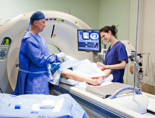
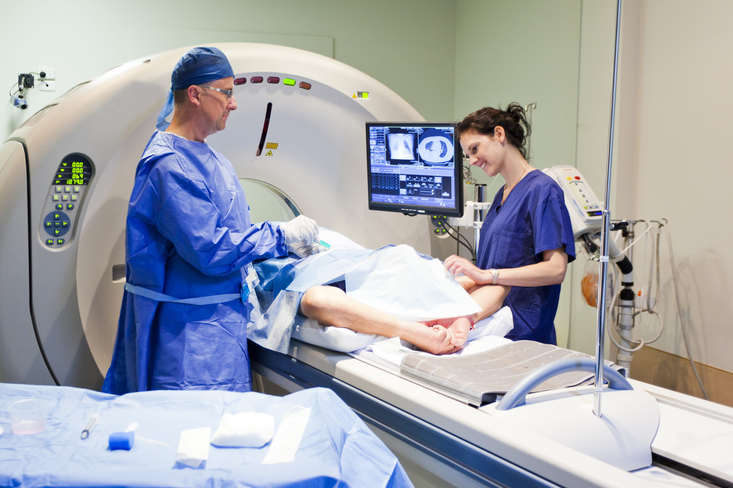
Innovations in medical and pharmaceutical imaging demand precision, speed, and reliability—and GaGe high-speed digitizers are rising to the challenge. From enhancing breast cancer detection and cardiovascular research to improving ultrasound clarity and gamma imaging portability, GaGe digitizers are powering breakthroughs across biomedical applications. This document highlights real-world case studies where GaGe technology has enabled advanced signal capture, leading to clearer diagnostics, deeper insights, and faster discoveries in healthcare and life sciences.
Precision Flow: Advancing Breast Health with 3D Imaging: Detect low-velocity micro-vascular blood flow with clarity & diagnostic confidence.

Challenge: Accurately mapping 3D blood flow in breast tissue is difficult due to the need to detect extremely low-velocity flow in tiny vessels, especially when the flow is nearly perpendicular to the imaging beam. Traditional imaging techniques often struggle to distinguish these subtle signals from surrounding tissue
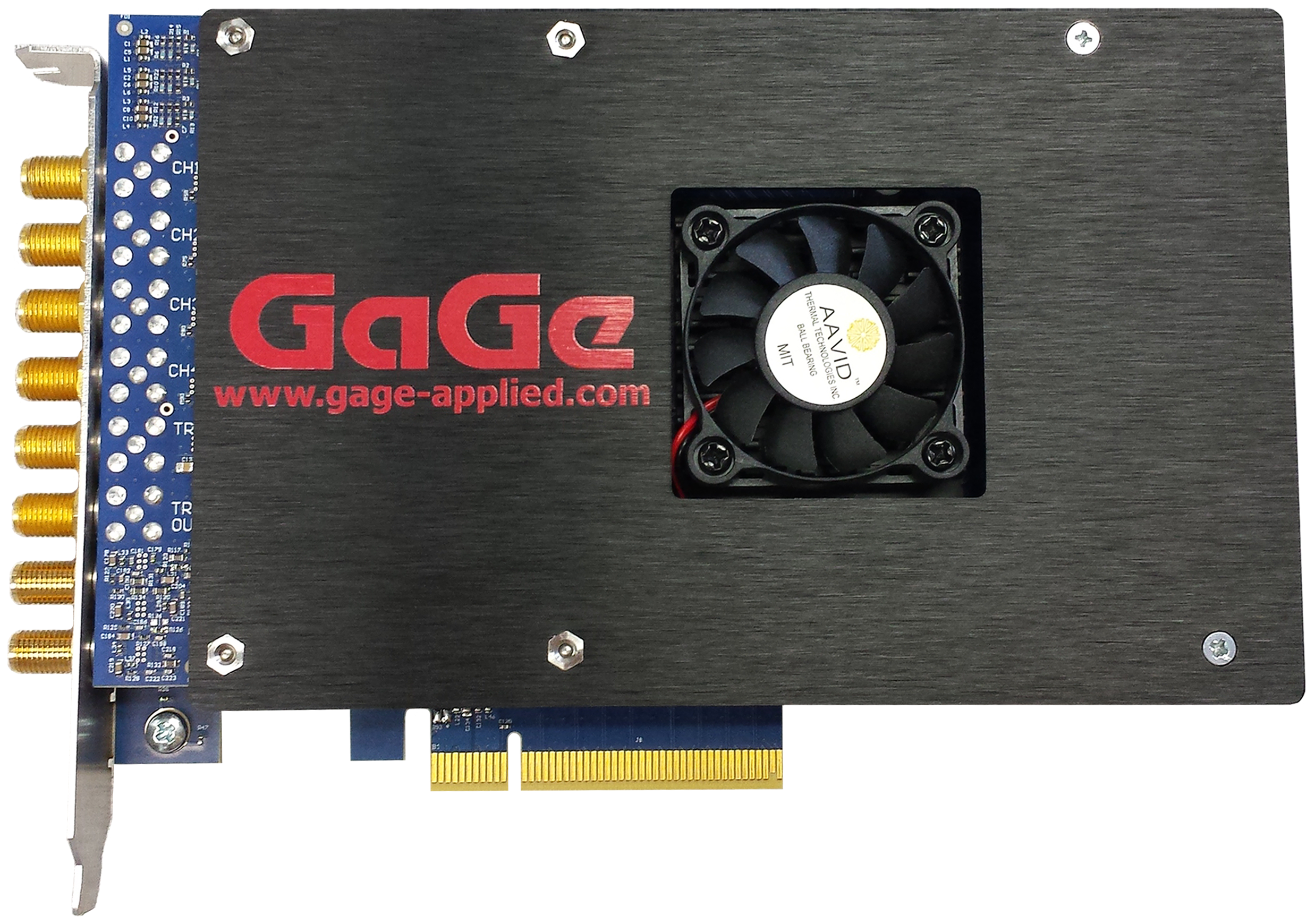
How the GaGe Digitizer was used: The research team used GaGe high-speed digitizers to capture wideband ultrasonic signals with high resolution and speed. This enabled the detection of low-velocity blood flow in tiny breast vessels by allowing long pulse integration and precise signal processing. The result: accurate 3D blood flow maps for earlier, more reliable diagnostics.
High Frequency Ultrasound: Advancing breast biopsy imaging.
 Challenge: Conventional ultrasound guidance during breast biopsies often fails to accurately detect micro-calcifications—small but critical indicators of early-stage breast cancer. These limitations can result in dangerous false negatives, putting patients at serious risk.
How the GaGe Digitizer was used: Researchers embedded a 64-element ultrasonic transducer array directly into the biopsy needle tip. Operating at 60 MHz, the high-resolution signals from each element were routed through a 64-to-1 multiplexer and digitized at 1 GS/s using a GaGe high-speed digitizer. This advanced configuration enables precise, real-time tracking of needle position—dramatically improving the accuracy of biopsies targeting micro-calcifications.
Tissue Clarity, Amplified: Solving imaging ambiguity with dual-color acousto-optics.
Challenge: Quantitative tissue imaging is limited by poor contrast and spectral ambiguity, making it hard to distinguish subtle differences in biological structures. This study introduces a two-color absorption interpolation technique using acoustooptic technology to enhance spectral accuracy.
Extraordinary Aspects of the Application: GaGe high-speed digitizer cards captured ultrafast acoustic signals with exceptional resolution and timing precision. Their performance enabled accurate reconstruction of dual-wavelength absorption data, providing sharper images and more reliable tissue characterization for advanced medical imaging applications.
Portable Precision: Solving cryogenic gamma imaging limits in SPECT and PET scans.
 Challenge: High-resolution gamma-ray detection is critical in medical imaging applications like SPECT and PET. However, traditional germanium detectors require cryogenic cooling, making them bulky, costly, and impractical for portable or bedside diagnostics. This study explores room-temperature alternatives like CdZnTe (CZT) detectors, which promise compact, clinical-friendly solutions—but demand exceptional signal fidelity.
How GaGe Digitizers were used: GaGe high-speed digitizers provide the precision and speed needed to capture and analyze fast gamma-ray signals from these detectors, enabling accurate, real-time imaging without liquid nitrogen. The result: advanced, mobile gamma imaging systems ready for modern medical environments.
Sharper Scans, Lower Noise Medical Imaging: Tackling FDML laser noise for sharper, more accurate diagnostics.
 Challenge: Medical imaging techniques like Optical Coherence Tomography (OCT) rely on FDML lasers for fast, detailed cross-sectional scans of tissues—essential in ophthalmology, cardiology, and oncology. However, FDML lasers often suffer from wavelength jitter and spectral noise, degrading image clarity and diagnostic accuracy. This research addresses these challenges by improving laser stability and spectral fidelity.
How the GaGe Digitizer was used: GaGe high-speed digitizers were instrumental in capturing ultrafast interferometric signals with high precision, allowing researchers to measure and correct spectral instabilities. A 12-bit, 200 MS/s Gage card acquires the signal from a 100MHz bandwidth photo receiver that is used to monitor laser power. The results are used to compensate for power fluctuations in the optical interferometer signal that is used to characterize the laser. The result: cleaner, sharper OCT images that empower clinicians with more accurate and reliable diagnostic information.
High Definition Diagnostics: Elevating medical ultrasound with high-frequency, high-speed signal capture.

Challenge: Conventional ultrasound systems are limited by lower frequencies and slower signal acquisition, restricting image resolution—especially when visualizing small or complex tissue structures in medical diagnostics. This research explores how high-frequency transducers paired with rapid data capture can dramatically enhance imaging detail.
Extraordinary Aspects of the Application: The article investigates usage of high frequency coded excitations to improve the dynamic range of ultrasonic imaging systems. In contrast to a conventional ultrasonic pulse excitation, a complex ultrasonic signal is generated by an Arbitrary Waveform Generator (AWG). The received ultrasonic signal is digitized by a 12-bit Gage card at 200 MS/s. While a conventional ultrasonic signal could be analyzed exclusively in the analog domain, digitization is required for coded signals, which must be subjected to extensive Digital Signal Processing (DSP) to recover conventional ultrasonic parameters.
Imaging the Unseen in Biomedical Research: High-resolution ultrasound for small animal cardiovascular studies.

Challenge: In biomedical research, small animal models are vital for understanding cardiovascular diseases and testing new therapies. However, traditional ultrasound systems struggle to capture the rapid, fine-scale dynamics of tiny hearts, like those in mice. This study presents a cutting-edge, high-frequency ultrasound system optimized for small animal imaging.
Extraordinary Aspects of the Application: GaGe high-speed digitizers enabled real-time acquisition of ultra-fast, high-resolution data, revealing subtle cardiac structures and functions previously invisible. The result: a powerful imaging tool that enhances biomedical research and accelerates discoveries in human cardiovascular health.
Ultrasound Precision for Medicine: Enhancing biomedical imaging with PFDF-on-silicon transducer technology.

Challenge: High-resolution ultrasound is vital in medical fields such as ophthalmology, cardiology, and dermatology, but traditional transducer materials often struggle to deliver the sensitivity and clarity needed at higher frequencies. Variability in fabrication techniques can also result in inconsistent imaging performance. This research presents an innovative approach to fabricating PVDF-on-silicon transducers, enabling thinner, more uniform layers with superior acoustic properties.
How the GaGe Digitizer was used: GaGe high-speed digitizers were key to validating these improvements—capturing the fine, high-frequency ultrasonic signals needed to measure and optimize transducer performance. The result: sharper, more reliable imaging that supports advanced biomedical diagnostic that empower clinicians with more accurate and reliable diagnostic information.


