
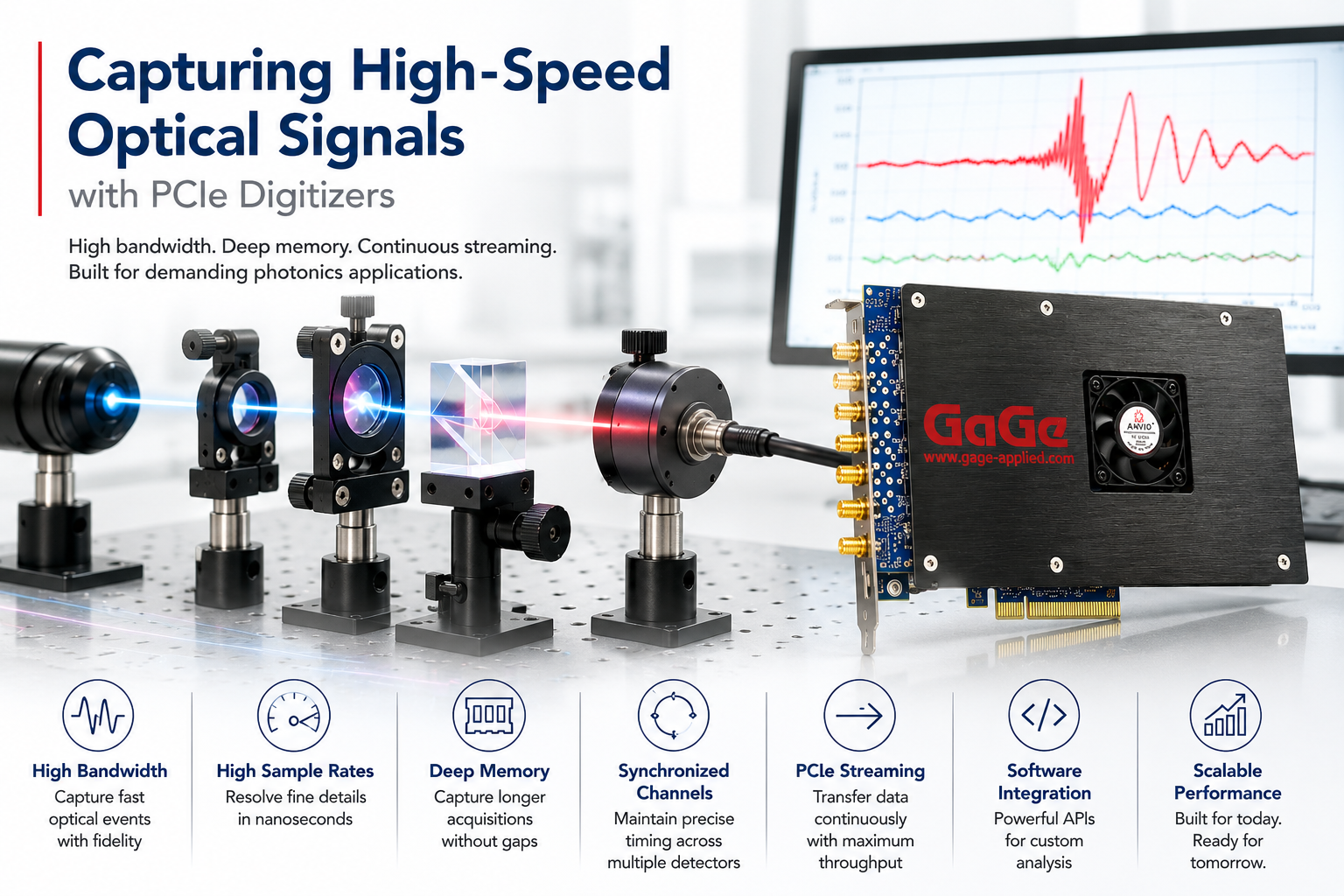
Introduction
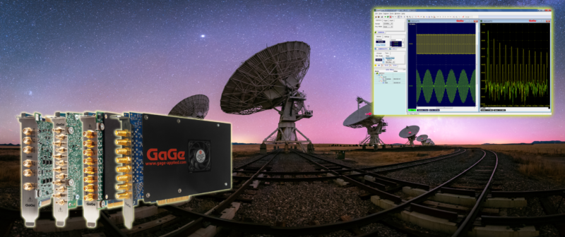
GaGe high-performance digitizers are playing a transformative role in advancing neurological diagnostics and brain research. GaGe digitizers provide the speed and precision needed for advanced brain imaging and neurological research. In focused ultrasound ther-apies, they enable real-time monitoring for opening the blood-brain barrier to deliver drugs safely and precisely. Their high-speed data capture also supports EEG, MEG, and brain-computer interface applications, offering accurate signal acquisition for diagnostics and therapeutic development. GaGe technology empowers deeper insights into brain function and next-generation neurotherapies.
Early Detection of Ovarian Cancer: Three-In-One Imaging Breakthrough for Early Detection of a Silent Killer

Challenge: Ovarian cancer is notoriously difficult to detect in its early stages. Often dubbed a “silent killer,” it presents few symptoms until it has already progressed to an advanced stage—when treatment options are limited and survival rates drop significantly. Traditional diagnostic tools lack the resolution, imaging depth, or contrast needed to identify malignancies before they spread. As a result, there’s an urgent need for a more effective diagnostic solution that can detect ovarian cancer earlier and more reliably.
How the GaGe Digitizer was used: The solution is a three-in-one endoscopic probe that combines optical coherence tomography (OCT), photoacoustic imaging (PAI), and ultrasound (US) to provide comprehensive, real-time imaging for early ovarian cancer detection. High-speed GaGe digitizer cards enable synchronized data acquisition from all three modalities, ensuring detailed and accurate diagnostic information from a single procedure.
Cracking Cataracts: Precision Lens Hardness Mapping with Ultrasonic Imaging

The article is a feasibility study of using high-frequency ultrasonic Nakagami imaging for characterizing the cataract lens in vitro.
Challenge: Cataracts harden the eye’s lens over time, and successful treatment often depends on knowing just how hard the lens has become. Unfortunately, current preoperative techniques do not provide a reliable, quantitative way to assess lens hardness. This forces surgeons to estimate, which can lead to inefficient energy use during ultrasound treatment and increase the risk of complications.
How GaGe Digitizers were used: Researchers used a 14-bit, 200 MS/s Gage digitizer to capture precise waveforms from a 35 MHz ultrasonic transducer aimed at pig eye lenses. These high-resolution signals enabled Nakagami statistical analysis to accurately characterize lens hardness, demonstrating a potential path to more tailored and efficient cataract treatment.
Pulse Check: Smart Imaging for Early Artery Disease Detection

Challenge: Arterial stiffness is a leading indicator of cardiovascular disease, yet traditional imaging methods struggle to detect subtle variations in vessel wall properties, especially in early stages. Identifying localized stiffness or inhomogeneity — particularly in small or complex arteries — requires high-resolution imaging and precise signal acquisition, which can be difficult to achieve in both phantom models and live subjects.
How GaGe Digitizers were used: Researchers employed Adaptive Pulse Wave Imaging (PWI) to monitor pressure wave propagation and vessel wall stiffness using both 5 MHz and 30 MHz ultrasonic transducers. The transducers were translated across arterial phantoms and mouse aortas to map spatial inhomogeneities in stiffness. A 14-bit, 200 MS/s Gage digitizer was used to capture high-speed, high-fidelity signals from the ultrasound probes, enabling accurate tracking of wall motion and stiffness in real time. This approach demonstrates a powerful, automated method for early detection of arterial abnormalities and cardiovascular risk.
Seeing Through the Needle: High-Res Imaging for Accurate Breast Cancer Detection

Challenge: Breast biopsies play a critical role in detecting early signs of cancer, such as microcalcifications. However, conventional ultrasonic imaging used to guide biopsy needles often lacks the precision needed to accurately target small or ambiguous lesions. This limited imaging resolution can lead to false negatives, delaying diagnosis and potentially compromising patient outcomes.
How GaGe Digitizers were used: To enhance biopsy accuracy, researchers developed a novel approach using a miniature 74 MHz ultrasonic transducer embedded directly into the biopsy needle. This high-frequency sensor provides real-time, localized imaging to supplement standard ultrasound guidance. A Gage digitizer card was used to acquire the rapid, high-resolution waveforms from the transducer, enabling precise visualization of tissue structures at the needle tip. This integrated solution significantly improves targeting accuracy, reducing the risk of missed diagnoses.
Striking Gold: Photoacoustic Imaging for Stem Cell Monitoring

Challenge: Stem cell therapies lack reliable in-body tracking. Stem cells hold tremendous promise for healing a variety of diseases, but a major limitation remains: once injected, it’s difficult to track their location and behavior inside the body. Without a reliable method to monitor stem cells in real time, evaluating their effectiveness or guiding treatment becomes a significant challenge.
How the GaGe Digitizer was used: To overcome this issue, researchers explored tagging stem cells with gold nanocages, which can be detected using photoacoustic microscopy. Two in vitro tracking methods were tested: two-photon microscopy and photoacoustic imaging. For the photoacoustic approach, a 50 MHz ultrasonic transducer captured signals from the gold nanocages, and a 12-bit, 200 MS/s Gage digitizer amplified and digitized those signals with high precision. This setup offers a promising path toward real-time, non-invasive monitoring of stem cell therapies.


