Treadmill Power Glitch Monitoring
Treadmill Power Glitch Monitoring Customer Case A customer develops gym equipment such as treadmills. The requirement is for an inexpensive way to monitor voltage signals for long periods of time. The treadmills are controlled by microprocessors that drive the LED displays. The customer has observed that some units intermittently fail during testing, a condition known as a "glitch" or a "brown-out". A glitch can have many causes, one of which is a weak power supply. The customer needs to capture certain key signals when a glitch occurs. At that stage, based on the captured data, it can be determined [...]

Press Release: Vitrek Launches New Electrical Safety and Test Equipment Catalog
Vitrek Launches New Electrical Safety and Test Equipment Catalog Vitrek’s new product catalog features sales, application and technical information for the company’s extensive line of electrical safety and test equipment. […]
Fiber Optic Sensor Measures Very Small Movements in Piezo Stacks
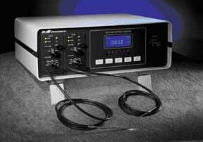
MTI Instruments, a global supplier of precision measurement solutions, has released an application note that explains how to measure very small movements in piezo devices that convert mechanical energy into electrical energy. Precision measurement of these dynamic, oscillatory motions is required for the research and development (R&D), testing, and troubleshooting of piezo ceramic materials, advanced ceramics that are used in actuators, sensors, and the power modules of ultrasonic welders. Piezo applications range from automotive sensors and medical devices to engineering projects and consumer products. MTI’s App Note explains how its MTI-2100 Fotonic sensor, when used in conjunction with a [...]

Capacitive Displacement Sensors: Working Principle & Precision Measurement Applications
Capacitive Displacement Sensors: Working Principle & Precision Measurement Applications Capacitive displacement sensors are precision non-contact measurement devices that utilize capacitance changes to determine position, distance, and thickness with exceptional accuracy. These capacitive distance sensors offer superior performance in applications requiring high-resolution measurements, making them essential tools across manufacturing, automation, and research industries. How Capacitive Displacement Sensors Work Capacitive Sensor Working Principle Capacitive displacement sensors operate on the fundamental principle of measuring capacitance variations between two conductive surfaces. The sensor probe acts as one plate of a parallel plate capacitor, while the [...]

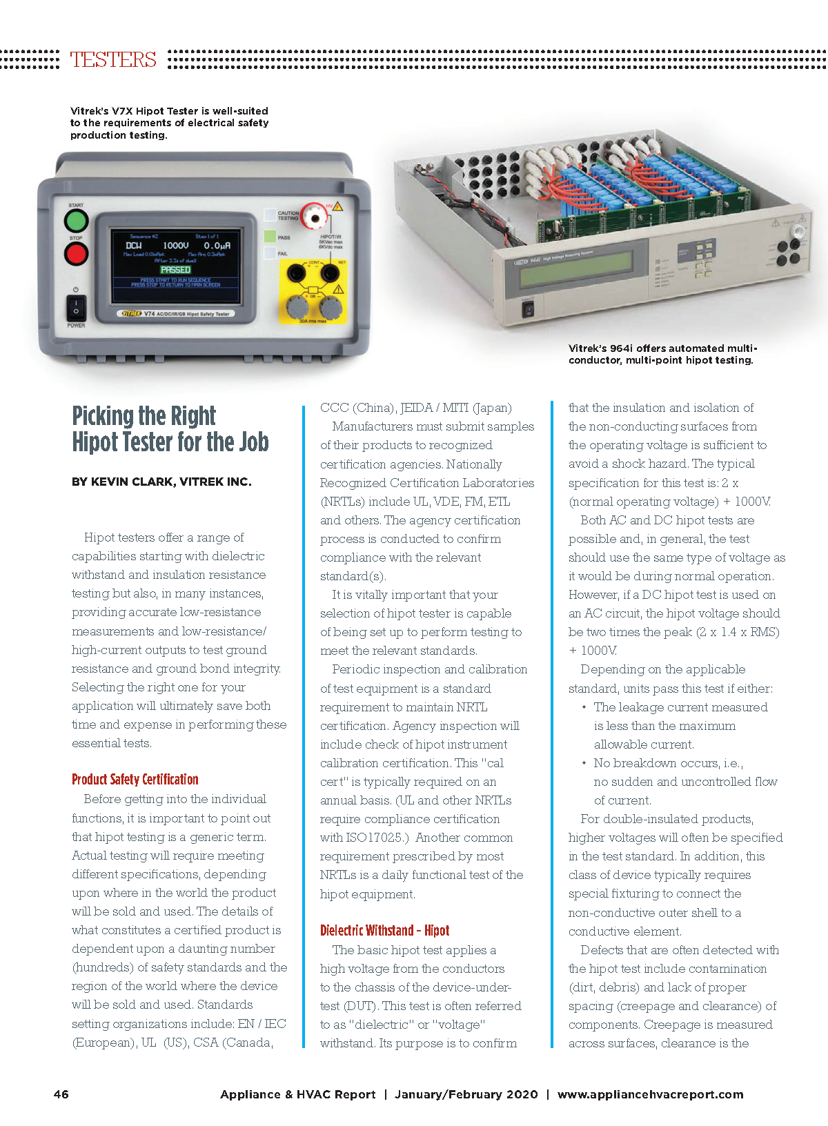
Vitrek featured in Appliance & HVAC Report
Click here to download a copy of this article featuring Vitrek Products! Click here to download a copy of this article featuring Vitrek Products!

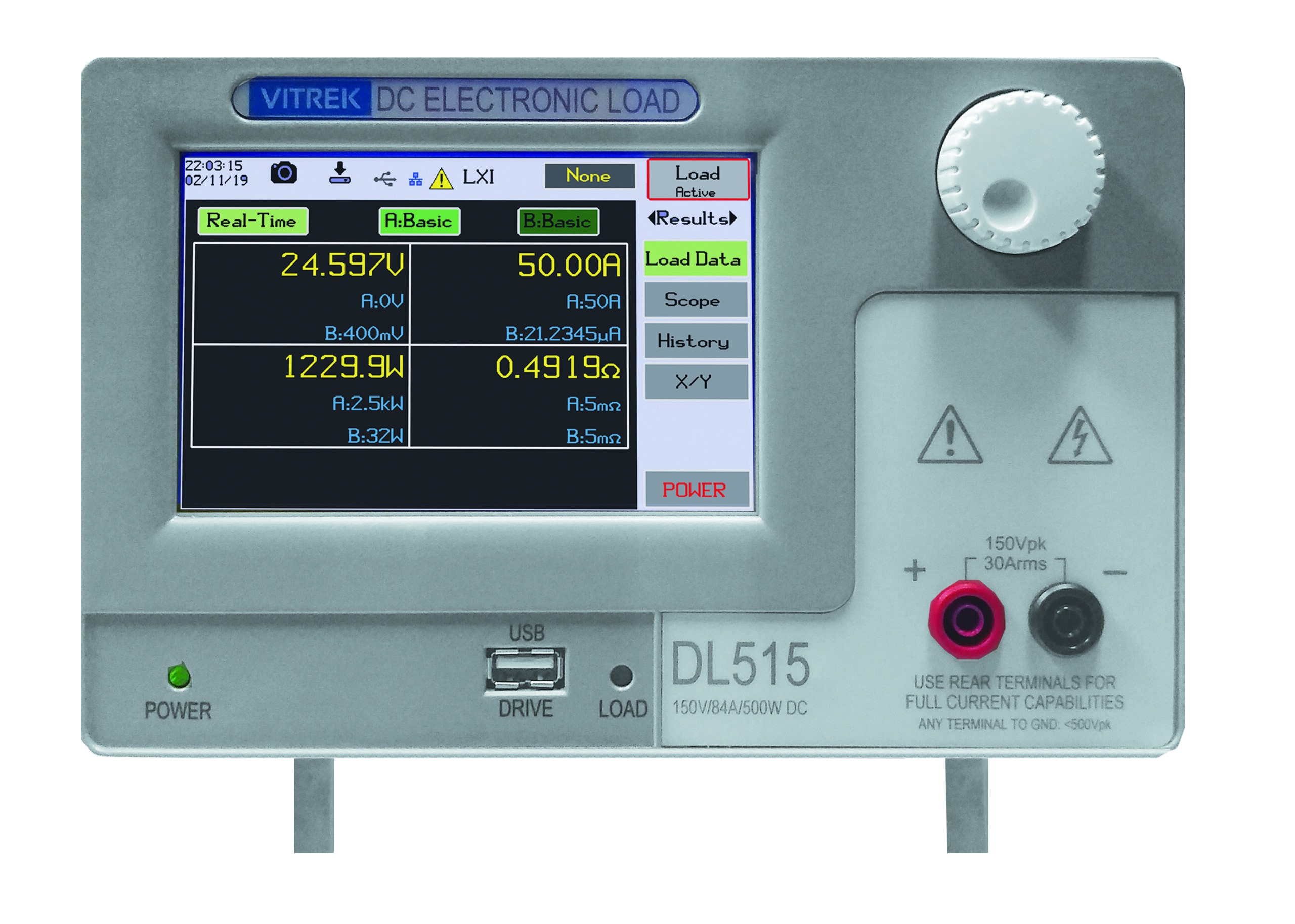
Press Release: Vitrek Launches New Programmable DC Load Product
Vitrek Launches Best-In-Class Programmable DC Load Product The Vitrek DL Series offers industry-leading performance in 125W-500W applications; supports best-in-class transient loading capability (from 0.1µW resolution to 14.5kW pulses); features high-speed pulse loading up to 100 kHz; boasts up to ten times better measurement accuracy. […]

Vitrek featured in Power Systems Design Magazine
Click here to download a copy of this article featuring Vitrek Products!

Vitrek featured in Design World Magazine
Click here to download a copy of this article featuring Vitrek Products!
 Position, Displacement, and Vibration Measurement with Precision, High Resolution, and Flexibility Gallery
Position, Displacement, and Vibration Measurement with Precision, High Resolution, and Flexibility
Application Notes-MTI, Brand-MTI, Industry-Aerospace, Industry-Automotive, Industry-Compliance Testing, Industry-Consumer Products, Industry-Government/Military, Industry-Manufacturing, Industry: Transportation, News-MTI-PBS, Products-MTI-Engine Balancing, Z-REPUB, z1
Position, Displacement, and Vibration Measurement with Precision, High Resolution, and Flexibility
The MTI-2100 Fotonic™ sensor uses advanced fiber optics and electronics to precisely measure position, displacement, and vibration. This high-resolution, non-contact metrology system supports a wide range of interchangeable fiber-optic probes and works with almost any surface, including metallic, composite, plastic, glass, ceramic, or liquid. Made in the USA by MTI Instruments, the MTI-2100 Fotonic™ features a modular design and supports application-specific requirements in R&D, quality, and process control. With its dual channel capabilities, the MTI-2100 Fotonic™ sensor allows users to make simultaneous measurements, such as for structure dynamics and modal analysis. All probe modules have two distinct operating ranges [...]
 How the Aviation Industry Solves Vibration and Balancing Challenges Gallery
How the Aviation Industry Solves Vibration and Balancing Challenges
Application Notes-MTI, Brand-MTI, Industry-Aerospace, Industry-Automotive, Industry-Compliance Testing, Industry-Consumer Products, Industry-Government/Military, Industry-Manufacturing, Industry: Transportation, News-MTI-PBS, Products-MTI-Engine Balancing, Z-REPUB, z1

How the Aviation Industry Solves Vibration and Balancing Challenges
The aviation industry spends significant amounts of time and money on problems caused by engine vibration. Imbalance, the most common cause of engine vibration, happens because of bird strikes and other impacts, but also because of natural wear and corrosion that redistribute mass. In turn, out-of-balance parts can result in cabin noise, metal fatigue, and cracked turbine, fan, or compressor components. In the most extreme cases, catastrophic engine failure may result MTI Instruments, a worldwide supplier of precision measurement solutions, makes equipment for monitoring vibration levels and balancing rotors. For maintenance, repair and overhaul (MRO) crews who want to [...]