
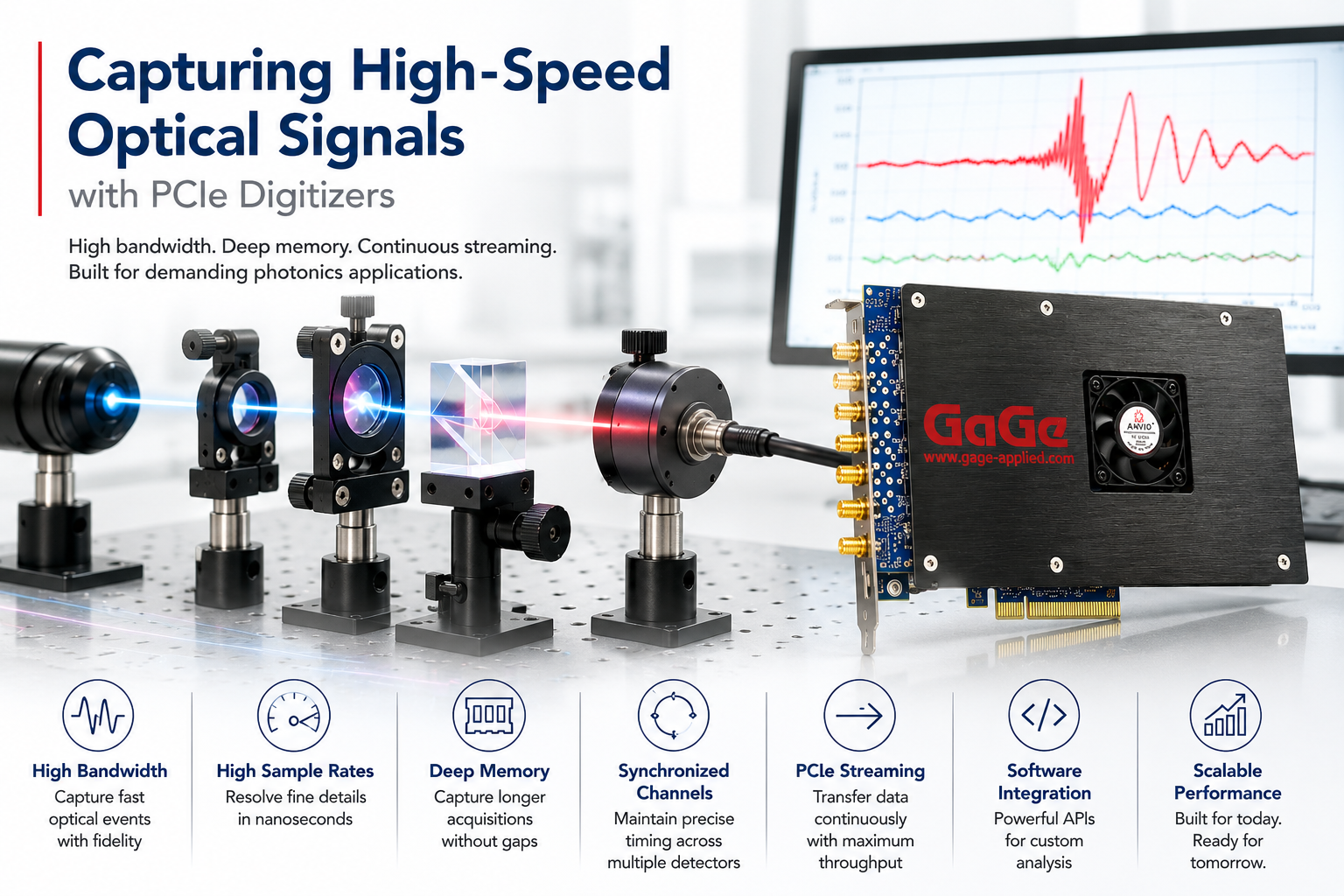
Introduction
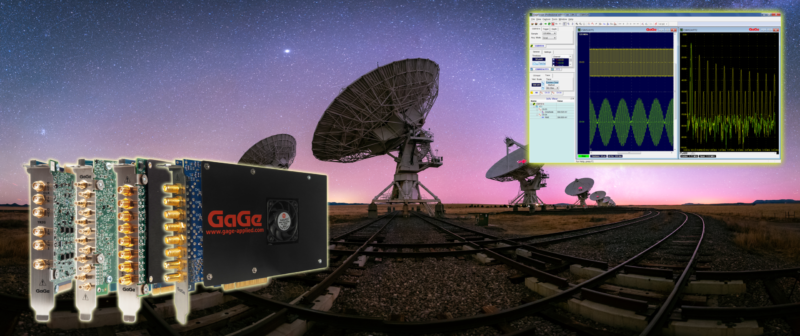
GaGe high-performance digitizers are playing a transformative role in advancing neurological diagnostics and brain research. GaGe digitizers provide the speed and precision needed for advanced brain imaging and neurological research. In focused ultrasound therapies, they enable real-time monitoring for opening the blood-brain barrier to deliver drugs safely and precisely. Their high-speed data capture also supports EEG, MEG, and brain-computer interface applications, offering accurate signal acquisition for diagnostics and therapeutic development. GaGe technology empowers deeper insights into brain function and next-generation neurotherapies.
Unlocking the Brain: Non-Invasive Method to Open the Blood-Brain Barrier using Ultrasound & Microbubbles

Challenge: The article studies usage of cavitating micro-bubbles to breach the Blood-Brain Barrier, which normally inhibits absorption of medication by the brain. A high power focused ultrasound transducer aimed at the brain is used to induce cavitation in microbubbles injected into the bloodstream. Monitoring a Passive Cavitation Detector (PCD) in contact with the brain to produce spectrograms has been shown to be a powerful technique to detect and characterize cavitation in mice brains. This study investigates usage of such a PCD to detect cavitation in monkey brains, where detection is more challenging because of the increased skull thickness.
How the GaGe Digitizer was used: The GaGe Digitizer was used to measure the electrical field induced by the TMS coils. The digitizer recorded the voltage differences captured by a dipole probe in the head model, allowing researchers to compute the electrical field strength and distribution.
Breaking Barriers: Targeted Brain Delivery Using Focused Ultrasound
The article investigates usage of cavitating micro-bubbles to breach the Blood-Brain Barrier and aid absorption of medication by the brain.
Challenge: The goal is to induce cavitation in microbubbles injected into the bloodstream. This induction is achieved by aiming high power focused ultrasound transducer at the brain.
How GaGe Digitizers were used: A Gage CompuScope digitizer monitors the amplitude of the excitation of the focused transducer. The resulting CompuScope data are used to estimate the absolute ultrasonic pressure, which allows study of its effect upon microbubble cavitation within mouse brains.
Neuroimmunomodulation: How Focused Ultrasound Enhances Brain Immune Defense
Challenge: Neuroimmunomodulation is a drug-free treatment in which the brain is irradiated by high-power focused ultrasound. The treatment can be effective for neurological diseases, such as Alzheimer’s.
The underlying mechanism of the treatment is poorly understood. This article experimentally compares three separate proposed mechanisms in mice brains. The irradiating high-power focused ultrasonic transducer contains a smaller low-power transducer that is used for alignment of the focused transducer and to monitor ultrasonic activity in the brain in some cases.
How GaGe Digitizers were used: Measurements of the absorption and scattering of infrared light can provide information, for example, about blood oxygenation and tissue composition. A Gage digitizer was used to monitor signals from two photodetectors – one that measures raw laser power and a second connected to the signal from the infrared interferometer that contains the sample. The Gage waveform data are then used to produce final infrared absorption and scattering measurements.
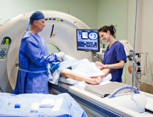
Brain Tissue Analysis: Making Brain Tissue Analysis Faster & Gentler with Ultrafast Lasers
In medical and brain research, it’s important to study tissues in great detail—right down to the molecules—without damaging the samples. Traditional methods can be rough and don’t always give a clear picture.
Challenge: Studying brain tissue at the molecular level is essential for advancing medical research, but traditional mass spectrometry methods can damage delicate samples and don’t provide the detailed images needed for 3D analysis.
How GaGe Digitizers were used: By combining ultrafast laser pulses with a high-speed GaGe Digitizer, researchers developed a powerful new imaging technique. This system gently removes and analyzes tiny layers of brain tissue, capturing precise signals in real time. The result: non-destructive, high-resolution 3D images that reveal both the structure and chemical makeup of brain samples—paving the way for better diagnostics and brain research.
Reaching Deep: Enhanced Brain Stimulation with Advanced H-Coils

The application refers to advancements in transcranial magnetic stimulation (TMS), a technique used in neuroscience and neurology for non-invasive brain stimulation. TMS is widely used for research & treatment of neurological & psychiatric disorders.
Challenge: Traditional TMS coils, like the figure-8 coil, have limitations in stimulating deep brain regions without causing unwanted cortical stimulation. This restricts the effectiveness of TMS for treating conditions that involve deeper brain structures.
How the GaGe Digitizer was used: The GaGe Digitizer was used to measure the electrical field induced by the TMS coils. The digitizer recorded the voltage differences captured by a dipole probe in the head model, allowing researchers to compute the electrical field strength and distribution.


